
Graphical Representation of Biomolecules
Antiparallel beta-sheet
This binder-protein conjugate contains several sections of antiparallel β-sheet, a common motif, represented in the cartoon mode by flat yellow arrows which indicate the N-to-C-to-C direction of the residues involved. To see the small molecule bound inside the protein, choose Trace from the Style menu followed by zoom in for closer view.
×
![]()
×
![]()
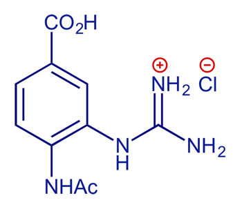
Structure-based inhibitors of influenza virus sialidase. A benzoic acid lead with novel interaction. [PDB structure code 1INF]
S. Singh, M. J. Jedrzejas, G. M. Air, M. Luo, W. G. Laver and W. J. Brouillette, J. Med. Chem., 1995, 38, 3217–3225